Facilities
Our facilities
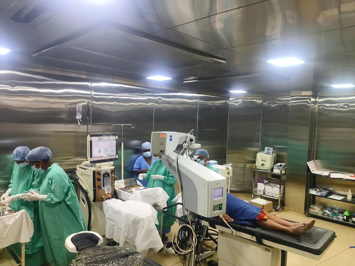
Operation theatre
Dedicated operation theatre for cataract and other eye surgery, with an operating microscope.
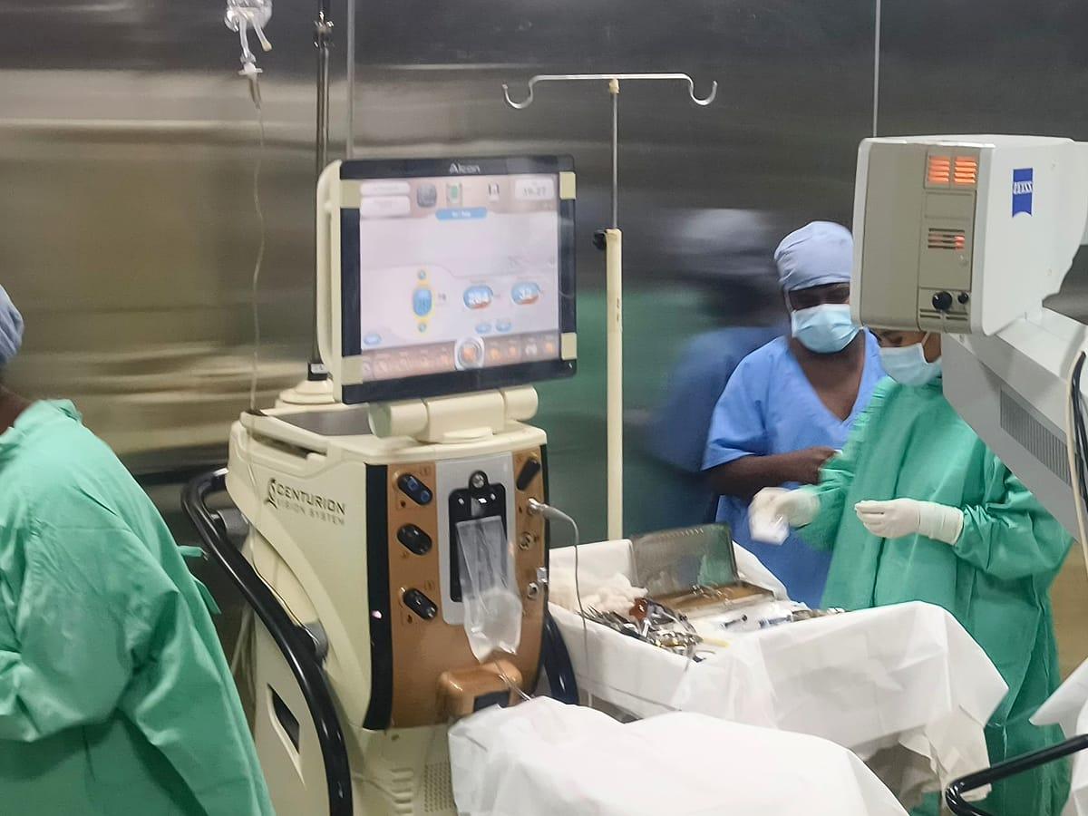
Phaco cataract system
Alcon Centurion phacoemulsification machine, used for small-incision cataract surgery.
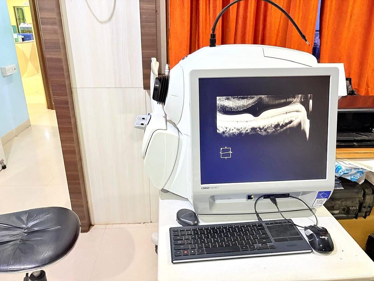
OCT scanner
Zeiss Cirrus HD-OCT 500 — detailed cross-section scans of the retina and optic nerve.
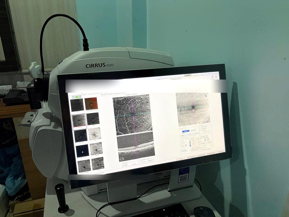
OCT angiography
Zeiss Cirrus 6000 — shows the blood vessels of the retina without a dye injection.
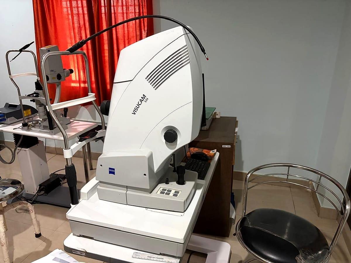
Fundus camera & FFA
Zeiss Visucam 524 — photographs of the retina, and fundus fluorescein angiography (FFA) of its blood vessels.
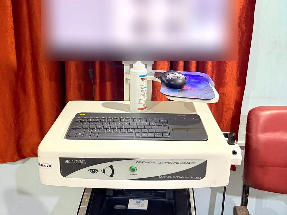
B-scan & UBM ultrasound
Appasamy Marvel scanner — ultrasound of the inside of the eye, and of the front of the eye in fine detail (UBM).
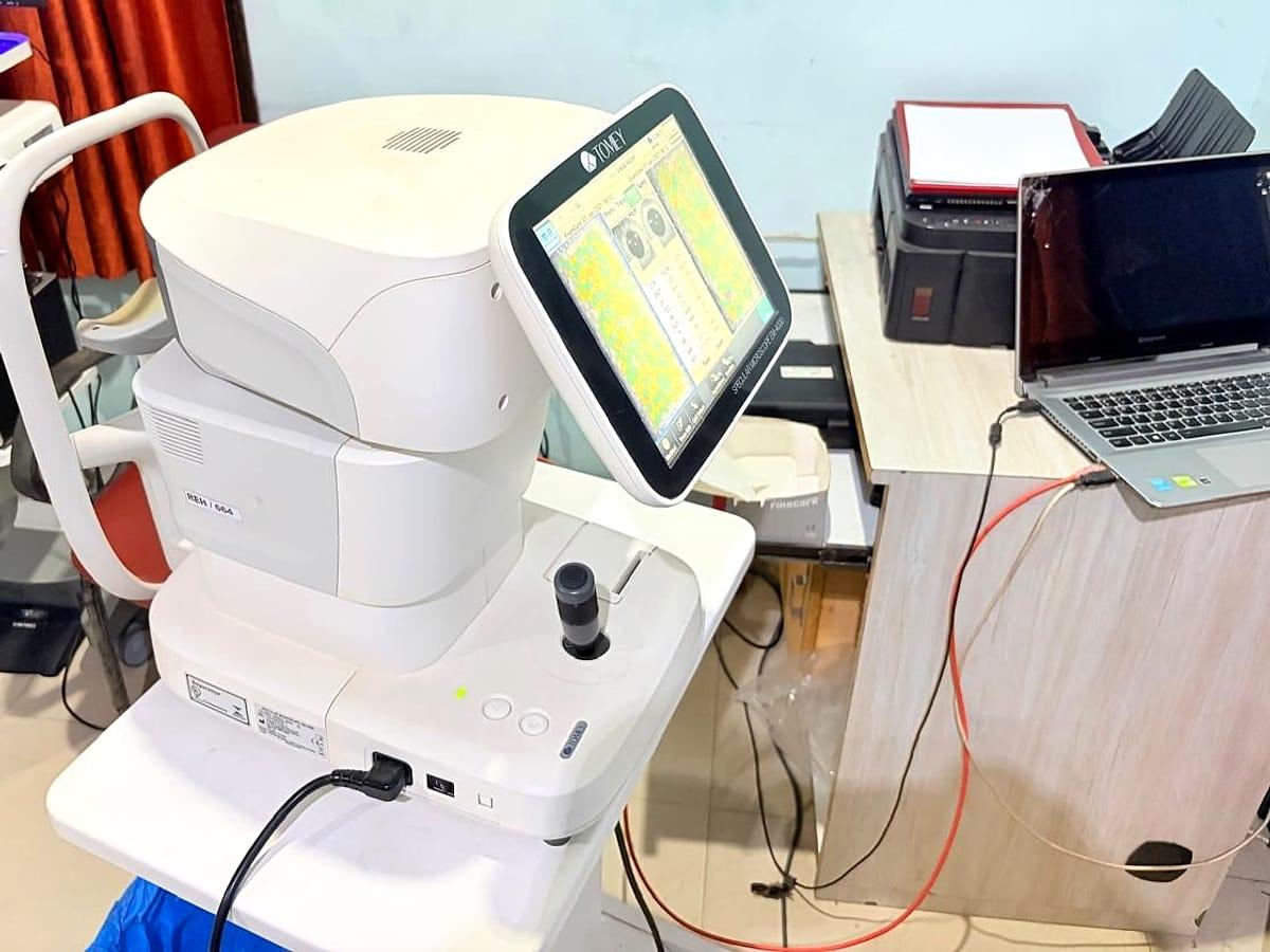
Specular microscope
Tomey specular microscope — counts the cells of the cornea's inner layer, e.g. before cataract surgery.
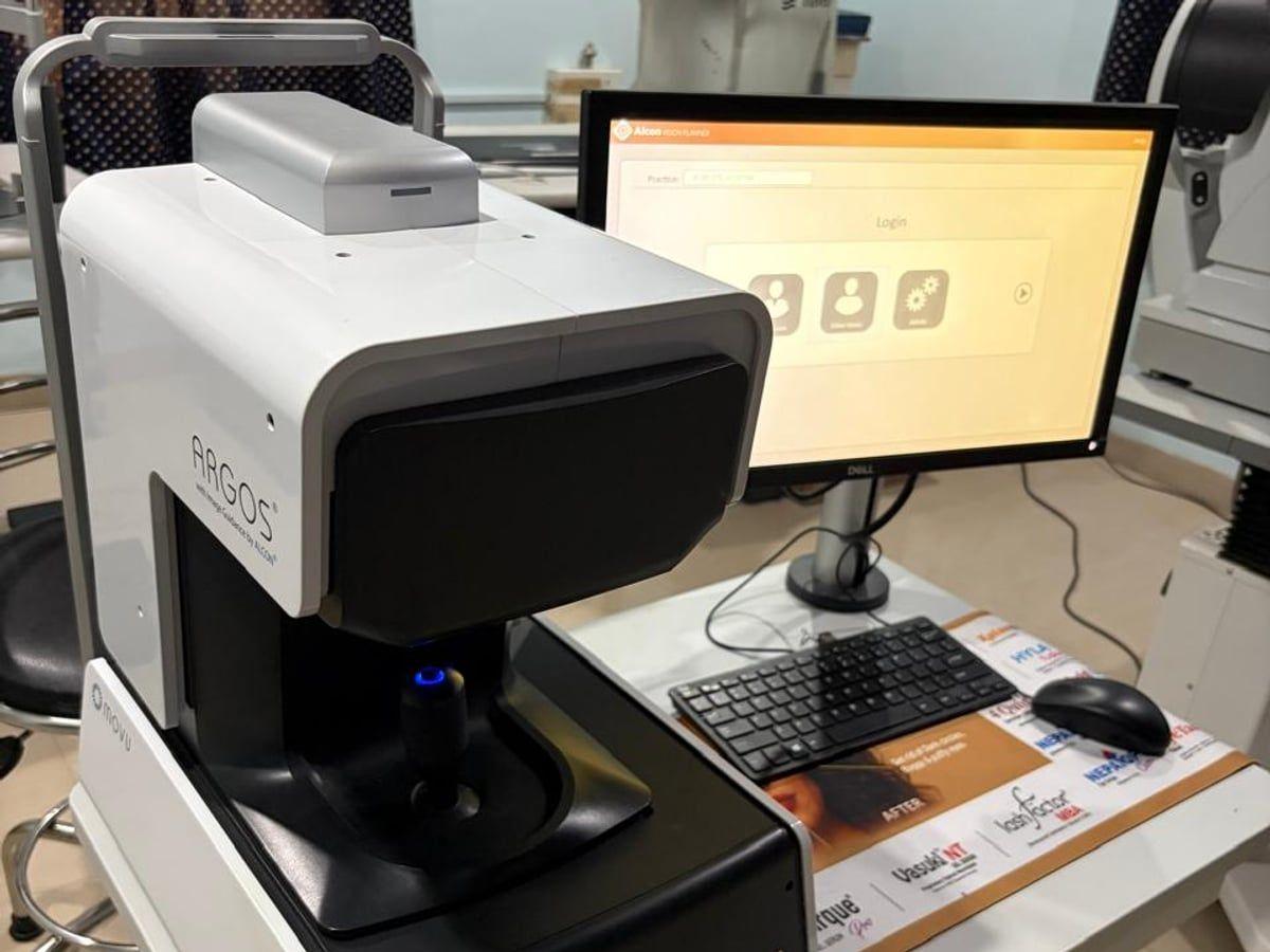
ARGOS optical biometer
Alcon ARGOS — measures the length and inner dimensions of the eye to calculate lens implant power for cataract surgery.
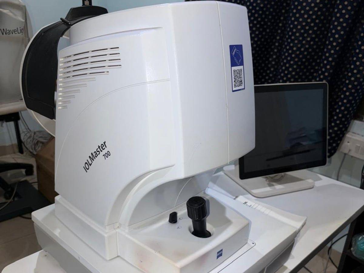
IOLMaster 700
Zeiss IOLMaster 700 — measures the eye without touching it, to choose the right lens implant before cataract surgery.
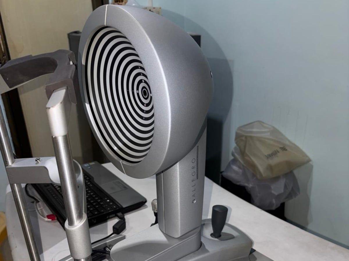
Corneal topographer
WaveLight Allegro Topolyzer — maps the curve of the cornea, to detect keratoconus and astigmatism and plan LASIK.
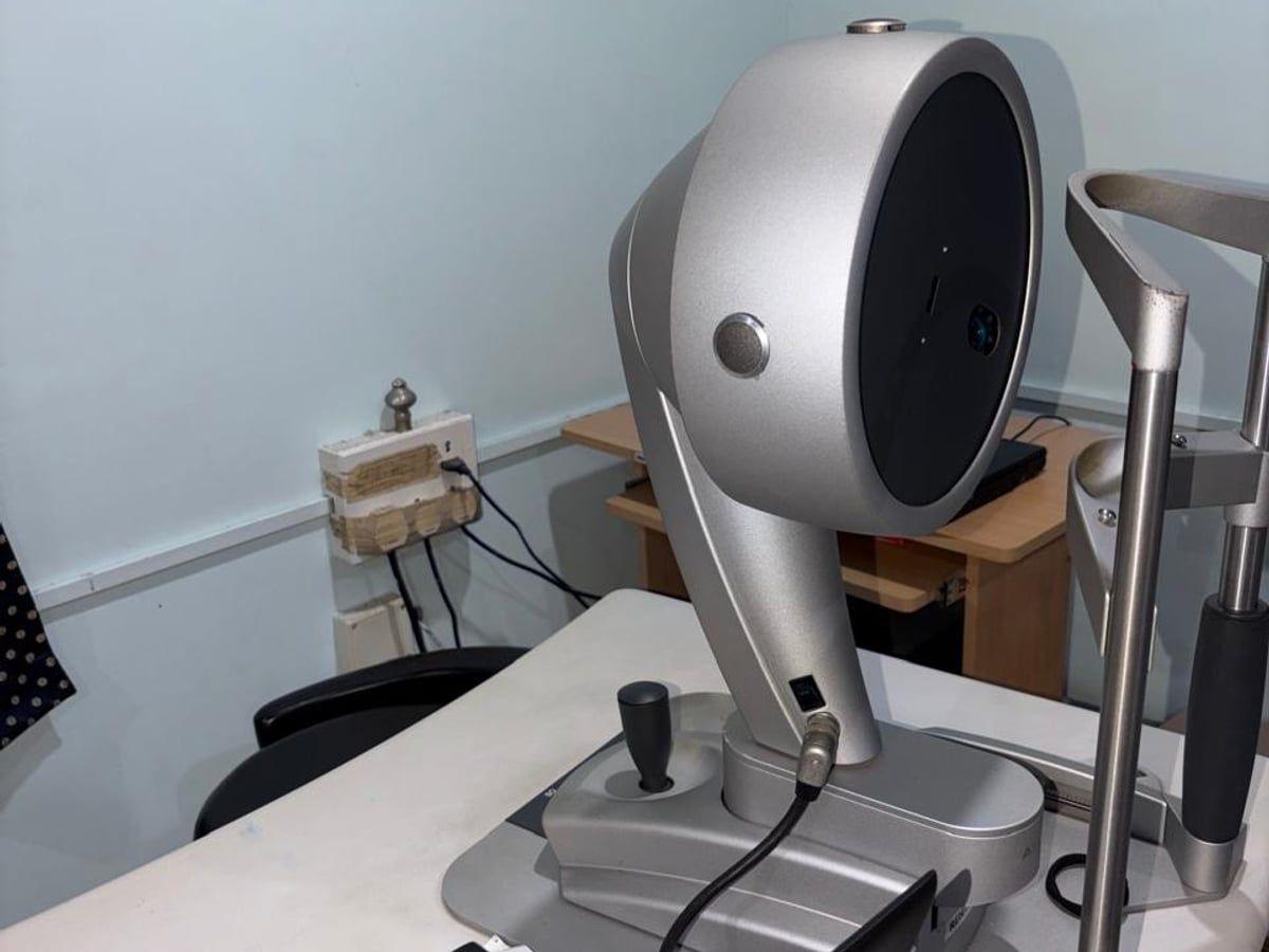
Corneal tomography
WaveLight Allegro Oculyzer — maps the shape and thickness of the cornea, used to plan LASIK and Contoura treatment.
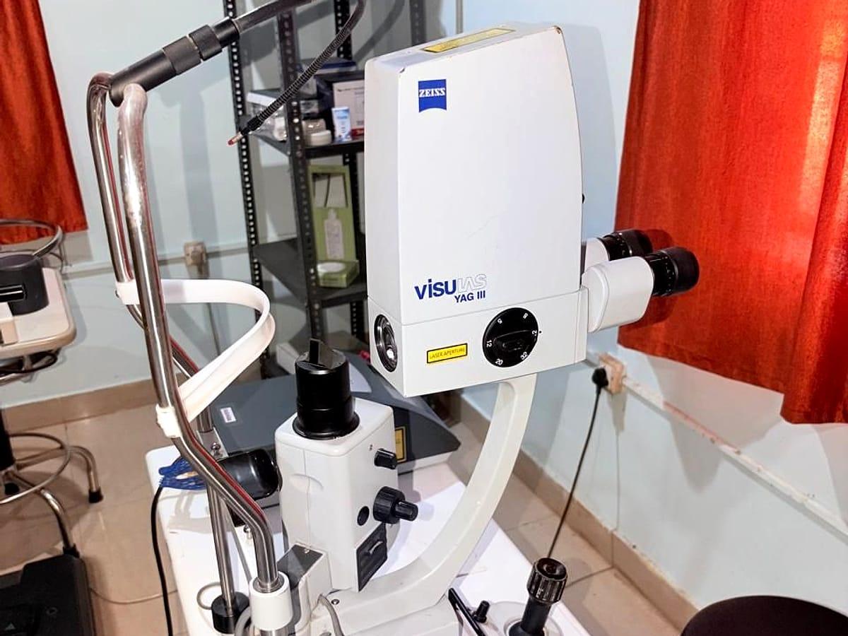
YAG laser
Zeiss Visulas YAG III — clears clouding behind a lens implant after cataract surgery and treats some glaucoma.
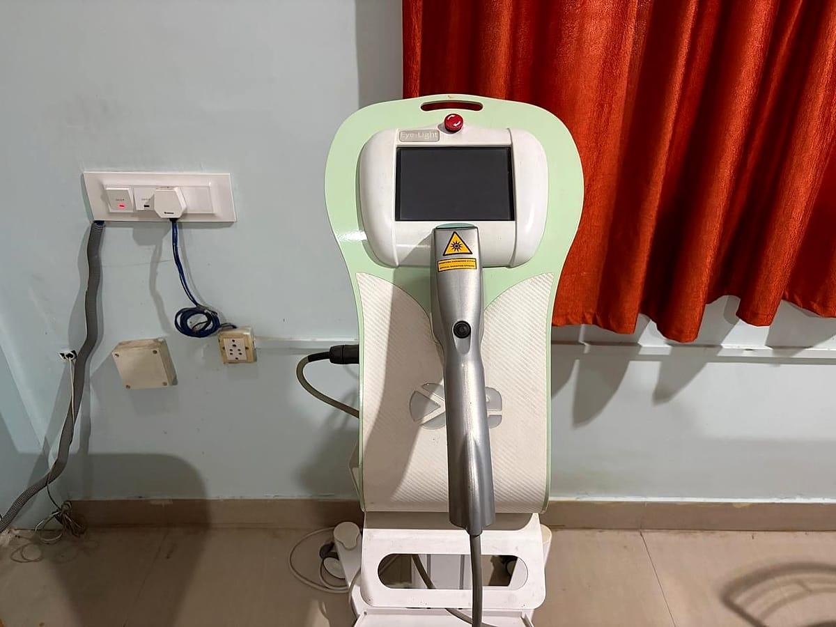
IPL dry eye therapy
Eye-Light intense pulsed light (IPL) treatment for dry eye caused by blocked oil glands in the eyelids.
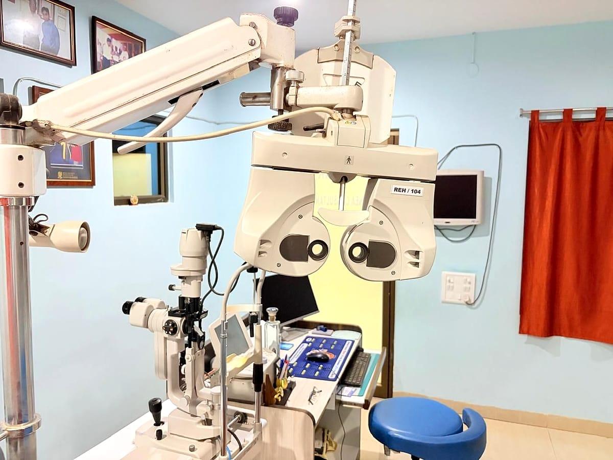
Consultation & refraction
Consultation rooms with slit lamp and phoropter for eye examinations and glasses power testing.
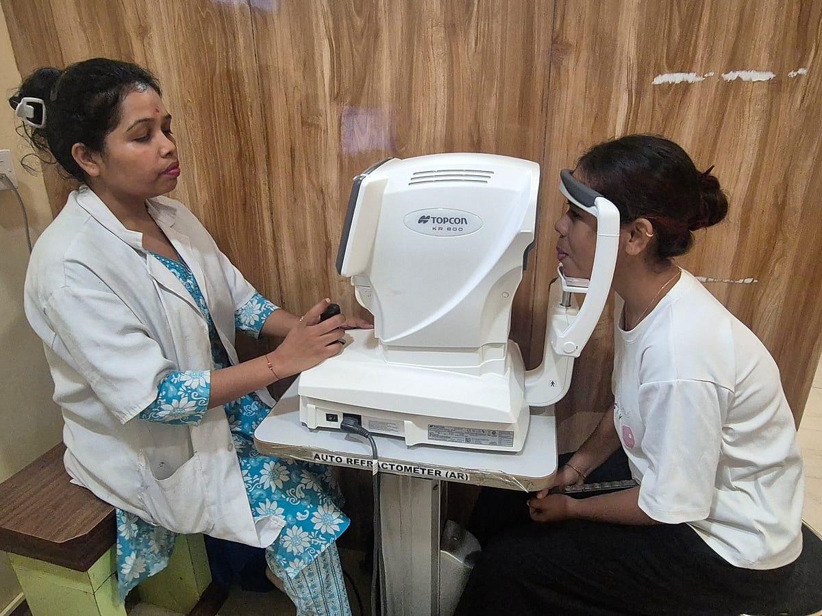
Computerised eye test
Topcon auto-refractometer — a quick computer reading of glasses power to start your eye test.
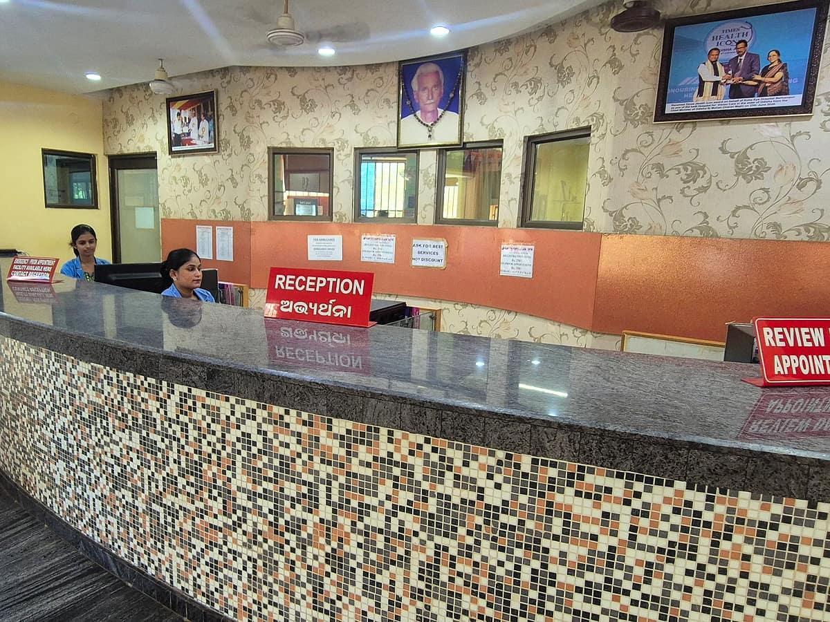
Reception
Front desk for registration, appointments and review dates.
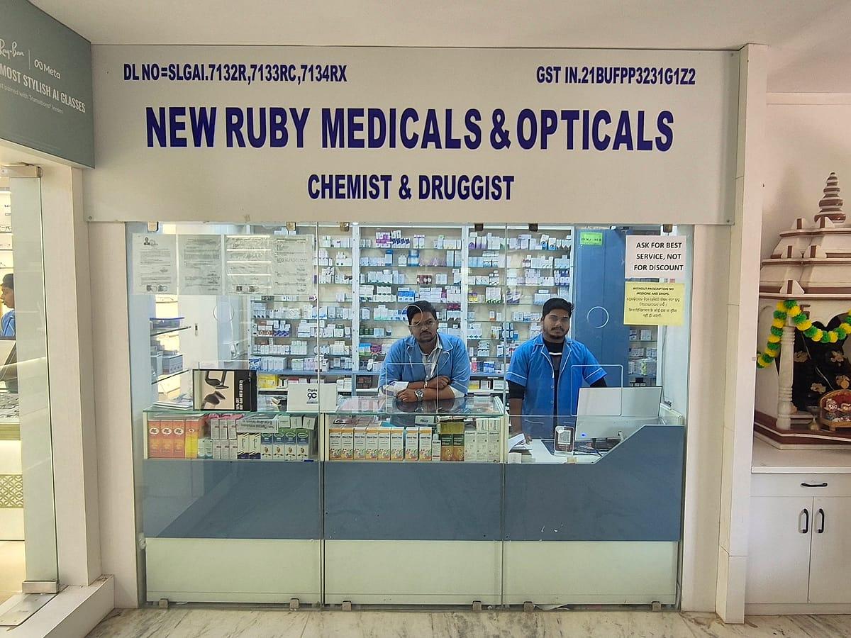
Pharmacy & optical counter
New Ruby Medicals & Opticals — medicines and spectacles on the premises.
LASIK & Contoura laser
Laser system for LASIK, Contoura LASIK and PRK vision correction.
Waiting area
Patient waiting area for patients and their families.
Instruments available
29 instruments for eye tests, scans, laser treatment and surgery — what each one is used for.
LASIK & vision correction
- LASIK EX-500 laser
Excimer laser that reshapes the cornea to correct glasses power.
- Contoura LASIK
LASIK planned from a detailed map of your own cornea.
Cornea mapping & measurements
Pentacam
Photographs the front of the eye to map the cornea's shape and thickness.
Oculyzer
Corneal mapping linked to the laser, used to plan LASIK and Contoura treatment.
Corneal topographer
Maps the curve of the cornea, e.g. to detect astigmatism or keratoconus.
AS-OCT
Scans the front part of the eye — cornea and drainage angle — without touching it.
Pachymeter & biometer
Measure the thickness of the cornea and the length of the eye.
IOL Master
Measures the eye without touching it, to choose the lens power for cataract surgery.
- ARGOS optical biometer
Measures the length and inner dimensions of the eye to calculate lens implant power.
Specular microscope
Counts the cells of the cornea's inner layer, e.g. before cataract surgery.
Cataract & eye surgery
- Infinity phaco system
Phacoemulsification machine for small-incision cataract surgery.
Operating microscope
Gives the surgeon a magnified, well-lit view of the eye during surgery.
Laser & cornea treatment
- YAG laser
Clears clouding behind a lens implant after cataract surgery, and treats some types of glaucoma.
- Green laser
Laser treatment of the retina, e.g. for diabetic retinopathy.
- LIO (laser indirect ophthalmoscope)
Delivers retina laser through a head-mounted scope, reaching the far edges of the retina.
- C3R (collagen cross-linking) machine
Strengthens a weak cornea in keratoconus with riboflavin drops and UV light.
IPL (intense pulsed light)
Treats dry eye caused by blocked oil glands in the eyelids.
Retina & glaucoma tests
- OCT
Cross-section scan of the retina and optic nerve, for macula, diabetic and glaucoma care.
OCT angiography
Shows the blood vessels of the retina without a dye injection.
FFA (fundus fluorescein angiography)
Photographs the blood vessels of the retina after a dye injection in the arm.
Fundus autofluorescence (FAF)
Non-invasive retinal imaging that checks the health of the retina's pigment layer.
Fundus camera
Colour photographs of the back of the eye, to record and follow changes.
Direct ophthalmoscope
Magnified examination of the optic disc, retina and retinal blood vessels during a routine eye check.
Indirect ophthalmoscope
Gives a wide view of the outer (peripheral) retina, used to check for retinal tears or detachment.
B-scan & UBM
Ultrasound of the eye when the inside can't be seen directly (e.g. behind a dense cataract); UBM shows the front of the eye in fine detail.
- Perimeter
Tests your side (peripheral) vision — important in glaucoma.
Non-contact tonometer (NCT)
Measures eye pressure with a gentle puff of air.
Eye tests & glasses power
Refraction unit
Workstation for testing glasses power and checking vision.
Autorefractometer
Gives a quick computer reading of glasses power to start the eye test.
CGHS, cashless & insurance
Bring your card or policy details to the front desk and our staff will help with the paperwork.
CGHS empanelled
Ruby Eye Hospital is a CGHS-empanelled eye centre, and cashless treatment is available. Please bring your CGHS card — the front desk will guide you through the paperwork.
Book an eye check-up
Request an appointment online and the hospital will call you to confirm, or call or WhatsApp us directly.