What it is
Retinal vein occlusion occurs when a vein carrying blood away from the retina, the light-sensitive layer at the back of the eye, becomes blocked. This can cause bleeding (retinal haemorrhage) and swelling within the retina, leading to blurred vision or sudden vision loss without eye pain. It is more common in people with high blood pressure, diabetes, high cholesterol or glaucoma.
Who it's for
Anyone with sudden blurring or loss of vision in one eye, without eye pain, should be seen urgently, as this can be a sign of retinal vein occlusion. People with high blood pressure, diabetes, high cholesterol or a history of glaucoma are at higher risk and should have regular eye checks.
What to expect
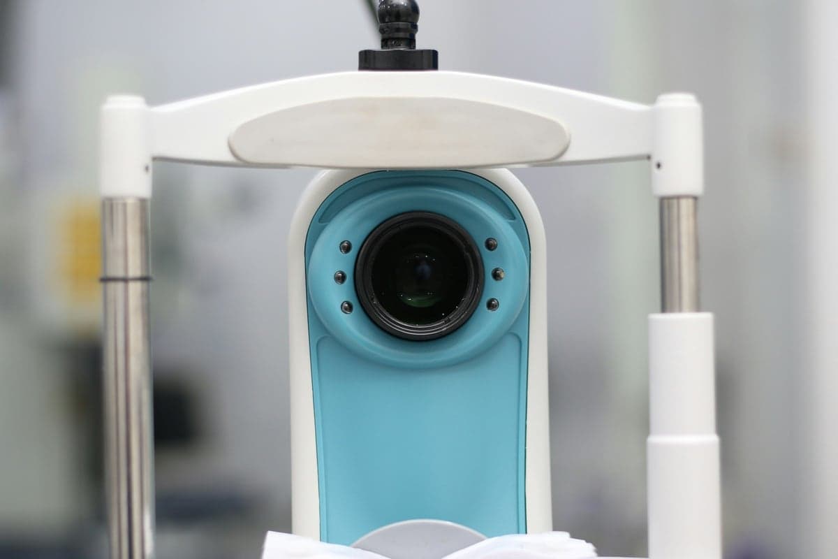
The retina is examined after dilating drops, usually with an OCT scan to check for swelling and sometimes a fundus fluorescein angiography (FFA) scan to assess blood flow. Treatment depends on severity and may include eye injections to reduce swelling, laser treatment in selected cases, and monitoring, alongside management of blood pressure, diabetes and cholesterol with your physician.
Recovery
Vision recovery varies depending on how much of the retina was affected and how soon treatment began. Many people need a series of visits and, if injections are used, repeat treatment guided by OCT scans. Keeping blood pressure, blood sugar and cholesterol under control reduces the risk of it affecting the other eye.
Frequently asked questions
Is retinal vein occlusion the same as a stroke in the eye?
It is sometimes described that way, since both involve a blocked blood vessel, but retinal vein occlusion affects the eye only, not the brain. It still needs prompt assessment.
Will my vision return to normal?
This depends on the extent of the blockage and how early it is treated. Some people recover good vision; others are left with some permanent blurring, which is why early assessment matters.
Medically reviewed by Dr. Sweta Patro